Module 03: Pattern Analysis Revised
Source: Dermoscopy Educational Course Authors: Harald Kittler, Cliff Rosendahl, and Alan Cameron
1. Learning Objectives
After completing this module, the learner should be able to:
- Define and identify the five basic geometric elements of dermoscopy (lines, pseudopods, circles, clods, dots) in clinical images.
- Recognize the six line patterns (reticular, branched, angulated, parallel, radial, curved) and the structureless pattern, and describe their formation from basic elements.
- Translate traditional metaphoric dermoscopic terminology (e.g., "pigment network," "blue-gray ovoid nests," "streaks") into objective geometric language.
- Describe the colors observed in dermoscopy and explain how melanin depth and other chromophores produce them.
- Apply the algorithmic approach of revised pattern analysis: describe patterns, assess symmetry, enumerate colors, and use clues to arrive at a diagnosis.
- Define chaos (asymmetry of structure or color) and explain its role as a screening criterion.
- List and describe the nine clues to malignancy used in the "Chaos and Clues" short algorithm.
- Differentiate between revised pattern analysis (specific diagnosis) and "Chaos and Clues" (biopsy-decision triage), understanding when each is appropriate.
2. Prerequisites
- Module 01: Introduction and Principles of Dermoscopy -- Equipment, polarized vs. nonpolarized dermoscopy, image capture techniques.
- Module 02: Histopathologic Correlations of Dermoscopic Structures -- Understanding the histologic basis for colors, networks, globules, dots, regression structures, and other dermoscopic features.
3. Key Concepts
Why Revised Pattern Analysis?
Traditional pattern analysis, while recognized as the most powerful tool for analyzing pigmented skin lesions by dermoscopy, evolved organically without an overarching logical framework. This led to:
- Opaque terminology -- Metaphoric language (e.g., "leaf-like areas," "moth-eaten border") that is subjective and difficult for beginners to apply consistently.
- Post-hoc description -- Descriptions sometimes constructed to fit a diagnosis rather than preceding analysis.
- Inconsistent criteria -- Lack of precise, repeatable definitions across observers.
Revised pattern analysis addresses these problems by:
- Using simple geometric terms instead of subjective metaphors.
- Requiring description before analysis -- observation must precede diagnosis.
- Establishing a standardized description language common to all diagnostic algorithms.
- Being applicable to every pigmented lesion (melanocytic and nonmelanocytic) on every anatomic site.
- Aiming for the most specific diagnosis possible, not merely "benign" vs. "malignant."
In 2015, the International Society of Dermoscopy (IDS) incorporated this descriptive terminology into its framework of standardized terms, providing a dictionary of both suitable metaphoric and descriptive terms.
4. Core Content
4.1 Description of the Method
Revised pattern analysis is a system based on terms defined clearly, precisely, and lucidly, applicable to every morphologic aspect of dermoscopy. Its fundamental principle is that description should always precede analysis. Although in practice description and analysis often proceed simultaneously, logically, description must come first. This precedence becomes critically important in the assessment of difficult lesions, where the clarity gained from a full formal description may lead to a diagnosis or, at minimum, ensures all relevant differential diagnoses are considered.
Description in this context is not a "photographic" impression of the lesion's appearance. Rather, it is the extraction of features relevant to diagnosis in the simplest possible terms.
Check Your Understanding
What are the five primary dermoscopic structural elements in the revised pattern analysis framework?
The five primary structural elements are lines (reticular, branched, parallel, radial, curved), dots, clods (globules), circles, and pseudopods. Structureless areas are also recognized as a sixth category.
4.2 Basic Elements
All patterns observed in dermoscopy are composed of five simple geometric elements:
| # | Element | Definition |
|---|---|---|
| (i) | Line | A two-dimensional continuous object with length greatly exceeding width, extending in one direction. |
| (ii) | Pseudopod | A line with a bulbous end. |
| (iii) | Circle | A curved line equidistant from a central point. |
| (iv) | Clod | Any well-circumscribed, solid object larger than a dot. Clods may take any shape. |
| (v) | Dot | An object too small to have a discernible shape. |
Vessel morphology is described using the same objective geometric terms as pigmented structures, with additional line types defined for vessels (as these are rarely seen in pigmented structures):
| Vessel Type | Description |
|---|---|
| Dots | Vessels appearing as dots |
| Clods | Vessels appearing as clods |
| Straight | Lines with no bend |
| Looped | Lines with one sharp bend |
| Curved | Lines with one gentle bend |
| Serpentine | Lines with multiple bends |
| Helical | Lines with multiple bends along a central axis |
| Coiled | Lines with multiple, tight bends |
4.3 Patterns Formed by Basic Elements
A pattern is built up of multiple repetitions of the same single basic element. There is also a pattern called "structureless" -- an area characterized by the absence of any basic elements, or at least with no basic element dominating.
Key rule: One or two repetitions of a basic element are not sufficient to form a pattern. A pattern must cover a significant part of the lesion (at least 25%) and be discernible by scanning the overall appearance.
Exceptions to the 25% rule:
- Any pseudopods or two or more radial lines are sufficient to constitute a pattern.
- Any reticular lines not due to collision with another lesion also constitute a pattern.
The Six Line Patterns
Lines form the most common and most specific patterns in dermoscopy:
| Line Pattern | Description |
|---|---|
| Reticular | Straight lines crossing each other at 90-degree angles, forming a grid. |
| Branched | Straight lines crossing each other at various (non-90-degree) angles. |
| Parallel | Straight lines that do not cross each other. |
| Radial | Straight lines that converge to the center of the lesion or to a central dot or clod. |
| Curved | Lines that bend (are not straight). |
| Angulated | Straight lines that do not intersect and meet at angles larger than 90 degrees, forming complete or incomplete polygonal shapes. |
Fundamental Principle of Term Application
The same term is applied to patterns composed of the same type of basic element, regardless of color or diagnosis. For example:
- "Brown globules" = brown clods
- "Red lacunes" = red clods
- "Blue ovoid nests" = blue clods
- "Cobblestones" = large polygonal clods
Variations in Patterns
Variations of basic patterns may be based on:
- Morphologic appearance: e.g., reticular lines may be thin or thick.
- Distribution of basic elements: e.g., on acral skin, parallel lines may lie on ridges, in furrows, or may cross ridges and furrows.
Pigmented skin lesions may consist of a single pattern or two or more patterns. Two or more patterns can be combined in a symmetric or asymmetric fashion. Two patterns can combine symmetrically in three ways (illustrated with reticular lines and dots):
- Dots at the periphery, reticular lines in the center.
- Dots in the center, reticular lines at the periphery.
- Dots scattered evenly on reticular lines.
All other combinations of two patterns will result in an asymmetric pattern.
Check Your Understanding
How do you differentiate between dots and clods on dermoscopy?
Dots are small round structures too small to discern any further morphology (typically less than 0.1 mm). Clods (globules) are larger round to oval structures where shape can be appreciated. The distinction is primarily one of size.
Key Takeaways
- The five basic dermoscopic elements are lines, dots, clods, circles, and pseudopods (plus structureless areas as a sixth category) -- all other terms can be translated into these.
- Lines forming a reticular pattern correspond to rete ridge pigmentation, while radial lines at the periphery (streaks/pseudopods) indicate active peripheral growth.
- Dots smaller than 0.1 mm reflect melanin granules or small nests, while clods larger than 0.1 mm represent larger nests or tumor islands.
4.4 Translation of Metaphoric Language
The following table translates traditional metaphoric dermoscopic terminology into simple geometric language based on the five basic elements:
| Metaphoric Language | Simple Geometric Terms |
|---|---|
| Pseudonetwork | Circles, brown, and confluent |
| Strawberry pattern | Circles, red |
| Grapes | Circles, small |
| Blue-gray ovoid nests | Clods, blue |
| Comedo-like openings | Clods, brown or orange (rarely black), and circles |
| Cobblestone pattern | Clods, large, brown or skin colored, polygonal |
| Milky red globules | Clods, pink and small |
| Red lacunes | Clods, red |
| Globules | Clods, small, round, or oval |
| Milia-like cysts | Dots or clods, white |
| Peppering | Dots, gray |
| Annular-granular pattern | Dots, gray and circles, gray |
| Stars in the sky | Dots, white |
| Streaks | Lines |
| Branched streaks | Lines, branched |
| Brain-like appearance | Lines, curved, and thick |
| Fat fingers | Lines, curved, and thick |
| Fissures and ridges | Lines, curved, and thick |
| Fingerprinting | Lines, curved, and thin (plus circles occasionally) |
| Fibrillar pattern | Lines, parallel, crossing fissures, and ridges |
| Peripheral streaks | Lines, radial |
| Radial streaming | Lines, radial, and segmental |
| Starburst pattern | Lines, radial or pseudopods, peripheral |
| Leaf-like areas | Lines, radial, connect to a common base |
| Spoke wheel-like area | Lines, radial, converging to a central dot or clod |
| Pigment network | Lines, reticular |
| Atypical pigment network | Lines, reticular, and thick |
| Broadened pigment network | Lines, reticular, and thick |
| Delicate pigment network | Lines, reticular, and thin |
| Inverse pigment network | Lines, reticular, white or gray |
| Reticular depigmentation | Lines, reticular, and white |
| Negative pigment network | Lines, reticular, white or gray |
| Crypts | Lines, thick, and curved in combination with clods |
| Chrysalis / crystalline structures | Lines, white |
| Moth-eaten border | Sharply demarcated, scalloped border |
| Regular blotch | Structureless zone, centric |
| Irregular blotch | Structureless zone, eccentric |
| Blue hue | Structureless zone, blue |
| Blue-white veil | Structureless zone, blue |
| Milky red areas | Structureless zone, pink |
| Scarlike depigmentation | Structureless zone, white |
| Central white patch | Structureless zone, white, central |
Clinical Scenario
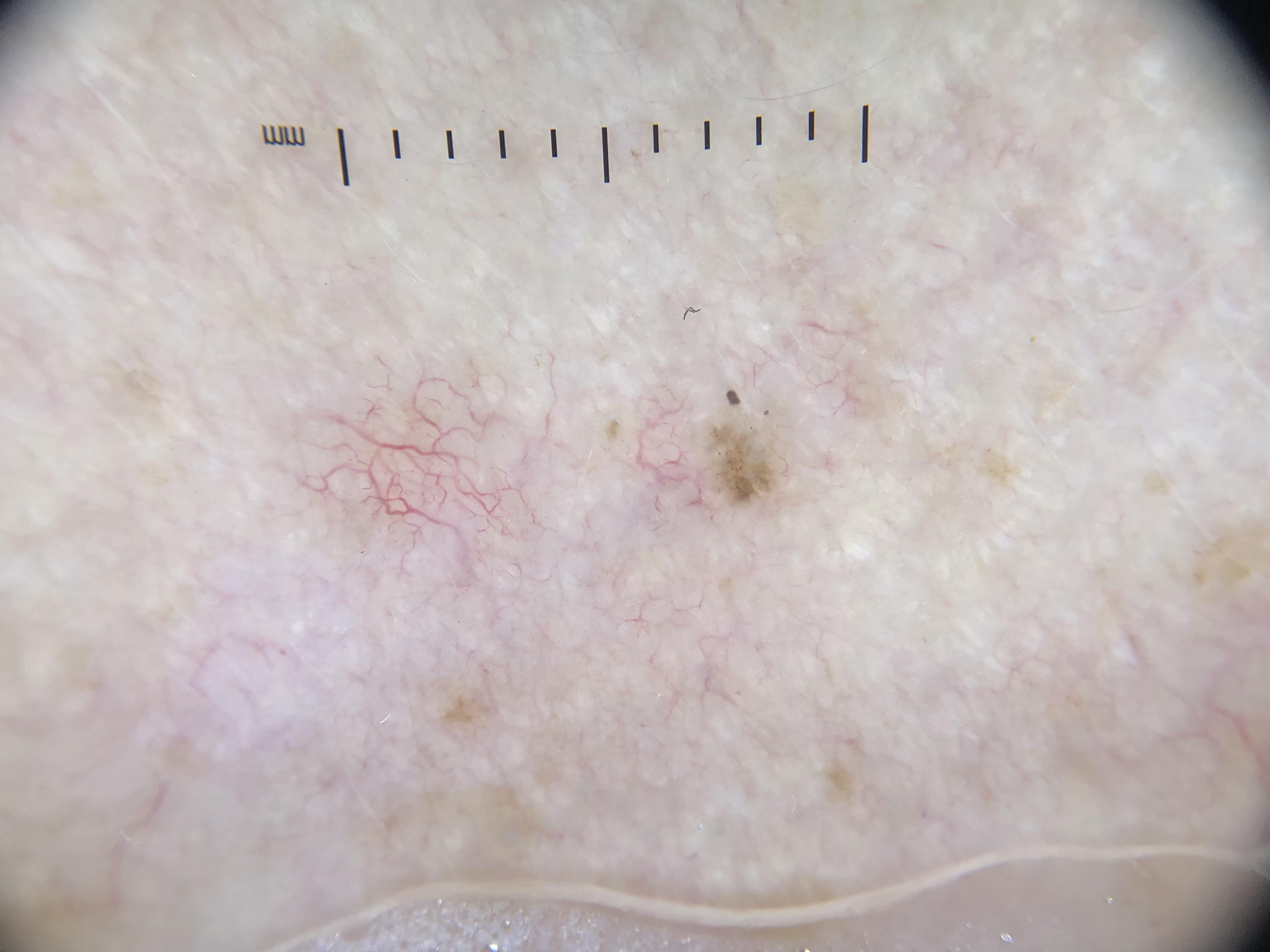
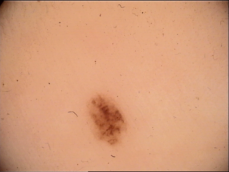
A 34-year-old woman presents with a 6 mm brown macule on the left thigh noticed 2 years ago. Dermoscopy reveals a single dominant pattern: thin lines crossing each other at 90-degree angles forming a grid (reticular lines), all of uniform thickness and light brown color. Small brown round structures (dots) are scattered on the lines. The overall appearance is symmetric.
What is your diagnosis and key dermoscopic findings?
Benign melanocytic nevus (junctional)
Using the revised pattern analysis framework, this lesion shows one dominant pattern (reticular lines, thin) with one color (light brown), making it symmetric by definition. The thin reticular lines correspond to a delicate pigment network -- the hallmark of benign melanocytic nevi with a junctional component. The dots on the lines are consistent with small melanocytic nests at the DEJ. A single symmetric pattern with uniform color strongly favors a benign diagnosis without need for further clue assessment.
4.5 Colors
Colors seen in dermoscopy are created by various combinations of:
- Keratin
- Melanin
- Blood (including serum in crusts)
- Collagen
- Foreign material
The color of melanin as seen by dermoscopy varies greatly depending on its localization in the epidermis or dermis:
| Melanin Location | Color Seen |
|---|---|
| Stratum corneum | Black |
| Superficial epidermis | Dark brown |
| Deep epidermis / dermoepidermal junction | Light brown |
| Papillary dermis | Gray |
| Deep dermis | Blue |
Other materials show less variation than melanin. Variation in color description between dermoscopists is minimized by:
- Not describing shades of brown, gray, and other colors.
- Classifying the number of colors only as either one or more than one color.
- When relevant, classifying colors only as being predominantly those of melanin or predominantly not those of melanin.
Check Your Understanding
What is a pseudopod, and what is its diagnostic significance?
A pseudopod is a bulbous projection connected to a pigmented lesion at its periphery, resembling a finger-like extension. Pseudopods indicate radial growth and are associated with melanoma when distributed asymmetrically, or with benign Spitz/Reed nevi when distributed symmetrically around the entire periphery.
Key Takeaways
- Metaphoric dermoscopy terms can be systematically translated: "pigment network" = reticular lines, "globules" = clods, "peppering" = gray dots, "crystalline structures" = shiny white lines.
- This standardized terminology reduces ambiguity and allows consistent communication across different dermoscopy schools of thought.
- Pattern recognition should always precede algorithmic analysis to ensure no structures are overlooked.
4.6 An Algorithmic Approach Using Patterns, Colors, and Clues
Revised pattern analysis offers a flexible framework for constructing a personal algorithm to derive as specific a diagnosis as possible for all lesions (benign or malignant, melanocytic or nonmelanocytic). It is based on the interpretation of patterns, colors, and clues.
Diagnosis follows a defined series of simple steps in logical sequence:
Step-by-Step Algorithm
Step 1 -- Describe the pattern(s): Determine the number of patterns. For analytic purposes, only two options are considered: one pattern or more than one pattern. This decision is rarely ambiguous.
Step 2a -- If ONE pattern:
- Characterize by color(s).
- A lesion with one pattern and one color is, by definition, symmetrical.
- If more than one color, assess symmetry.
- Formulate differential diagnosis based on pattern and color(s).
Step 2b -- If MORE THAN ONE pattern:
- Assess the combination for symmetry or asymmetry.
- The higher the number of patterns, the more unlikely the lesion displays symmetry.
- Analysis proceeds on the basis of the most specific pattern in the lesion, not the most prominent.
- Patterns are listed in order from most specific to least specific:
Lines > Pseudopods > Circles > Clods > Dots > Structureless
(most specific) (least specific)
- Therefore: lesions with lines are assessed by the differential diagnosis of lines. Only a lesion lacking lines is analyzed by pseudopods, and so on.
- A lesion that combines structureless with any other pattern is analyzed according to the other pattern (structureless is the least specific).
Step 3 -- Assess for clues (if needed):
- Assessment of patterns and colors may lead to a single diagnosis, but may also lead to a narrow differential.
- In that case, the lesion is examined for clues to resolve the differential.
- A clue is a feature too localized to constitute a pattern but nonetheless favoring one diagnosis over another.
- Sometimes a pattern may also constitute a clue (e.g., a structureless eccentric zone is both a pattern and a clue to melanoma).
General principle: More weight should be given to the overall pattern than to any single clue. The most difficult part of dermoscopy is correctly assigning weight to clues, particularly avoiding overvaluation of an unreliable or misleading clue. This is the role of experience.
Decision Flow
Lesion under examination
 |
 v
Describe pattern(s) and color(s)
 |
 v
 +-----+-----+
 | |
 v v
One pattern More than one pattern
 | |
 v v
Color(s)? Symmetry assessment
 | |
 v v
Symmetry? Analyze by MOST SPECIFIC pattern
 | (Lines > Pseudopods > Circles > Clods > Dots > Structureless)
 v |
Formulate v
differential Formulate differential
diagnosis diagnosis
 | |
 +------+------+
 |
 v
 Diagnosis reached?
 | |
 Yes No
 | |
 v v
 Done Examine for CLUES
 |
 v
 Resolve differential
4.7 A Short Algorithm Based on Pattern Analysis: "Chaos and Clues"
The authors recognize the need for a simple diagnostic system based on pattern analysis. "Chaos and Clues" simplifies the goal to identifying lesions requiring biopsy rather than reaching a specific diagnosis.
Key features:
- Applicable to every pigmented lesion.
- Does not require a decision as to whether a lesion is melanocytic.
- Detects pigmented malignancy with accuracy comparable to earlier systems.
- Tested on consecutive pigmented lesions, including significant proportions of benign and malignant nonmelanocytic lesions.
Key Takeaways
- Six dermoscopic colors (brown, black, gray, blue, white, red) each reflect specific chromophore depths and types in the skin.
- Multiple colors in a single lesion increase the probability of malignancy because they indicate architectural complexity across skin layers.
- The presence of blue and white together (blue-white veil) strongly suggests either melanoma or Reed nevus.
4.8 How the System Is Applied
Before applying "Chaos and Clues," a lesion of concern is assessed for pattern recognition morphology of any of the five common benign lesions:
- Nevus
- Benign keratinocytic lesion (solar lentigo, seborrheic keratosis, etc.)
- Dermatofibroma
- Hemangioma
- Sebaceous gland hyperplasia
If the lesion can be identified as one of these, the algorithm is not needed. If none can be identified, the algorithmic method is applied in two steps:
Step 1: Assess for CHAOS (asymmetry of structure or color)
 |
 +----+----+
 | |
 v v
 No chaos Chaos present
 | |
 v v
 BENIGN Step 2: Search for CLUES
 (stop) |
 v
 Clue(s) found?
 | |
 Yes No
 | |
 v v
 BIOPSY BENIGN
 indicated (stop)
Clinical Scenario
A 61-year-old man presents with a 12 mm irregular dark lesion on the upper back that has changed in color over the past 4 months. Dermoscopy reveals more than one pattern: thick reticular lines (thicker than the spaces they surround) in one area, an eccentric blue structureless zone in another, and segmental radial lines at the 7 o'clock position. At least four colors are present: brown, black, blue, and gray.
What is your diagnosis and key dermoscopic findings?
Melanoma (superficial spreading type)
Applying the pattern analysis algorithm: the lesion has more than one pattern, so we assess symmetry -- the combination is clearly asymmetric. Analysis proceeds by the most specific pattern (lines > pseudopods > circles > clods > dots > structureless). The thick reticular lines qualify as Clue 7 (lines reticular, thick -- thicker than the spaces they surround), produced by melanoma cells proliferating in the rete ridges. The eccentric blue structureless zone is Clue 1 (eccentric structureless area) and also Clue 2 (gray or blue structures). The segmental radial lines are Clue 4. Multiple clues in the presence of chaos mandate biopsy.
4.9 Chaos
Chaos is defined as the presence of dermoscopic asymmetry of structure or color.
- The shape of the lesion is not relevant.
- Any color other than skin color at the edge of a lesion (such as white) should be regarded as part of the lesion.
- Lesions without chaos are not analyzed further.
Three exceptions where biopsy is indicated even in lesions lacking chaos:
- Facial lesions with gray or blue structures.
- Acral lesions with parallel lines on the ridges.
- Any elevated, firm, and continuously growing lesion.
These exceptions should only be invoked in lesions that do NOT have the pattern recognition morphology of one of the five common benign lesions.
Important nuances:
- A benign seborrheic keratosis on the face may have central gray color, but symmetry, a uniform gradual border, and historical stability will confirm benign status.
- Many solar lentigines have pigmented circles, but a sharply defined scalloped border permits pattern recognition diagnosis, thus avoiding biopsy.
4.10 Clues to Malignancy
In "Chaos and Clues," a clue is a feature that, when present, indicates a lesion requires biopsy. One clue is sufficient. Both chaos and the clue can be produced by the same feature.
There are nine clues to pigmented malignancy (melanoma, pigmented BCC [pBCC], and pigmented Bowen disease [pBD]):
Clue 1: Eccentric Structureless Area
- Any color except skin color.
- An asymmetrically located structureless zone favors malignancy.
Clue 2: Gray or Blue Structures
- Gray dots may be seen in pBCC, pBD, and melanoma. They are a very common finding even in noninvasive melanomas, produced by free melanin or melanin in macrophages in the dermis.
- Gray circles occur in noninvasive facial melanomas.
- Dense deposition of melanin in the dermis causes blue clods or blue structureless areas in pBCC and invasive melanomas.
Clue 3: Black Dots or Clods, Peripheral
- Peripheral black dots and clods are generally produced by melanoma cells or nests, respectively, close to or at the level of the stratum corneum due to pagetoid spread.
- Black structures are rarely seen in pigmented BCC or pBD.
Clue 4: Lines, Radial or Pseudopods, Segmental
- Radial lines may be seen at any location in BCC.
- Pseudopods or radial lines when circumferential are a feature of Reed nevus, but if segmental, they are highly specific for melanoma.
Clue 5: Polymorphous Vessels
- Vessels are called polymorphous when more than one pattern of vessel is seen.
- Polymorphous vessels reflect disordered malignant growth pattern and possibly increased metabolic demand of actively growing tumor tissue.
- If vessels as dots are present, melanoma is more likely than BCC.
Clue 6: White Lines
- To qualify as a clue, white lines must be clearly lighter than perilesional skin.
Clue 7: Lines Reticular or Branched, Thick
- To be called thick, the lines must be thicker than the spaces they surround.
- This pattern is produced by melanoma cells proliferating in the rete ridges.
- Not seen in pBCC or pBD.
Clue 8: Angulated Lines
- Straight lines that do not intersect and meet at angles larger than 90 degrees, forming complete or incomplete polygonal shapes.
Clue 9: Lines, Parallel, Ridges (Acral Skin)
- This is a clue to malignancy even in the absence of chaos.
- Subcorneal hemorrhage also produces this pattern.
- Whether pigment lies on ridges or furrows is often easier to assess at the edges of a lesion.
Key Takeaways
- Chaos (asymmetry of structure and pattern) is the prerequisite for applying the Clues analysis; without chaos, a lesion is likely benign.
- The eight clues to malignancy include eccentric structureless areas, thick reticular or branched lines, gray or blue structures, black dots/clods in the periphery, polymorphous vessels, lines radial or pseudopods (segmental), and white lines.
- Any lesion with chaos plus at least one clue warrants biopsy or excision.
5. Pattern Recognition Reference Table
Table A: Basic Elements and Their Diagnostic Associations
| Basic Element | Appearance | Common Diagnostic Associations |
|---|---|---|
| Lines, reticular (thin) | Grid-like, thin lines crossing at 90 degrees | Benign melanocytic nevi (delicate pigment network) |
| Lines, reticular (thick) | Grid-like, thick lines (thicker than spaces) | Melanoma (atypical/broadened network), CLUE #7 |
| Lines, reticular (white/gray) | White or gray grid pattern | Melanoma (negative pigment network), dermatofibroma |
| Lines, branched | Lines crossing at various angles | Melanocytic lesions; if thick, CLUE #7 |
| Lines, curved (thin) | Thin, gently bending lines | Fingerprinting (solar lentigo, flat seborrheic keratosis) |
| Lines, curved (thick) | Thick, bending lines | Brain-like appearance, fat fingers, fissures/ridges (seborrheic keratosis) |
| Lines, parallel | Non-crossing straight lines | Acral lesions: furrows = benign; ridges = malignant (CLUE #9) |
| Lines, radial | Lines converging toward center | BCC (leaf-like areas, spoke-wheel areas); if segmental, CLUE #4 for melanoma |
| Lines, radial (peripheral/circumferential) | Radial lines around entire periphery | Starburst pattern (Reed/Spitz nevus) |
| Lines, angulated | Straight lines forming polygonal shapes (>90 degrees) | Melanoma on chronically sun-damaged skin, CLUE #8 |
| Lines, white | Lines clearly whiter than surrounding skin | Chrysalis/crystalline structures, CLUE #6 |
| Pseudopods | Lines with bulbous end | Starburst pattern; if segmental, CLUE #4 for melanoma |
| Circles, brown | Brown ring structures | Pseudonetwork (face), facial melanocytic lesions |
| Circles, red | Red ring structures | Strawberry pattern (actinic keratosis) |
| Circles, gray | Gray ring structures | Annular-granular pattern (facial melanoma), CLUE #2 |
| Circles, small | Small ring structures | Grapes pattern |
| Clods, blue | Blue solid objects | Blue-gray ovoid nests (BCC, blue nevus), CLUE #2 |
| Clods, brown/orange | Brown or orange solid objects with circles | Comedo-like openings (seborrheic keratosis) |
| Clods, large polygonal | Large, polygon-shaped solid objects | Cobblestone pattern (congenital/dermal nevus) |
| Clods, pink/small | Small, pinkish solid objects | Milky red globules (melanoma) |
| Clods, red | Red solid objects | Red lacunes (hemangioma, angiokeratoma) |
| Clods, small round/oval | Small, round/oval solid objects | Globules (nevi, melanoma) |
| Clods, black (peripheral) | Peripheral black solid objects | Pagetoid melanoma nests, CLUE #3 |
| Dots, gray | Gray point-like objects | Peppering/regression (melanoma, lichen planus-like keratosis), CLUE #2 |
| Dots, white | White point-like objects | Stars in the sky (milia-like cysts) |
| Dots or clods, white | White small objects | Milia-like cysts (seborrheic keratosis) |
| Structureless, centric | Central featureless zone | Regular blotch (benign nevus) |
| Structureless, eccentric | Eccentric featureless zone | Irregular blotch (melanoma), CLUE #1 |
| Structureless, blue | Blue featureless zone | Blue-white veil (invasive melanoma, BCC), CLUE #2 |
| Structureless, pink | Pink featureless zone | Milky red areas (melanoma) |
| Structureless, white (central) | Central white featureless zone | Central white patch (dermatofibroma) |
| Structureless, white | White featureless zone | Scarlike depigmentation (regression) |
Table B: Summary of Nine Clues to Malignancy
| Clue # | Name | Description | Malignancies |
|---|---|---|---|
| 1 | Eccentric structureless area | Any color except skin color, asymmetrically located | Melanoma, pBCC, pBD |
| 2 | Gray or blue structures | Gray dots, gray circles, blue clods, blue structureless areas | Melanoma, pBCC, pBD |
| 3 | Black dots or clods, peripheral | Due to pagetoid spread at/near stratum corneum | Melanoma (rare in BCC/pBD) |
| 4 | Lines radial or pseudopods, segmental | Circumferential = Reed nevus; segmental = highly specific for melanoma | Melanoma (segmental); BCC (any location) |
| 5 | Polymorphous vessels | More than one vessel pattern; dots favor melanoma | Melanoma, BCC |
| 6 | White lines | Clearly lighter than perilesional skin | Melanoma, BCC |
| 7 | Lines reticular or branched, thick | Thicker than spaces they surround; from melanocytes in rete ridges | Melanoma only (not pBCC or pBD) |
| 8 | Angulated lines | Straight, non-intersecting, >90-degree angles, polygonal shapes | Melanoma (especially sun-damaged skin) |
| 9 | Lines parallel, ridges (acral) | A clue even WITHOUT chaos; also seen in subcorneal hemorrhage | Acral melanoma |
6. Clinical Pearls
Description before diagnosis: Always describe what you see in geometric terms before attempting a diagnosis. This discipline prevents confirmation bias and ensures nothing is missed.
Most specific pattern governs analysis: When multiple patterns are present, analyze using the most specific pattern (lines > pseudopods > circles > clods > dots > structureless), NOT the most prominent one. This reduces diagnostic errors.
The 25% rule and its exceptions: A pattern must cover at least 25% of the lesion -- except for pseudopods (any), radial lines (two or more), and reticular lines (any not from collision), which always constitute a pattern regardless of area covered.
Chaos is a screening tool: Assessing for chaos (asymmetry of structure or color) requires minimal cognition and time with practice. Most benign lesions lack chaos, so this step efficiently triages the majority of lesions.
Segmental vs. circumferential radial lines: This distinction is critical. Circumferential radial lines/pseudopods suggest Reed/Spitz nevus (benign), while segmental radial lines/pseudopods are highly specific for melanoma.
Gray structures are high-yield: Gray dots are one of the most common findings even in noninvasive melanomas. On the face, gray circles are a key feature of lentigo maligna. Never dismiss gray structures.
Thick reticular lines are melanoma-specific among pigmented malignancies: When lines are thicker than the spaces they surround, this pattern is produced by melanoma cells in rete ridges and is NOT seen in pBCC or pBD.
Parallel ridge pattern is a clue even without chaos: On acral skin, parallel lines on ridges are sufficient for biopsy even in a symmetric-appearing lesion. Assess ridges vs. furrows at the lesion edges where it is often easiest to differentiate.
Three "no-chaos" exceptions: Remember that facial lesions with gray/blue structures, acral lesions with parallel ridge pattern, and any elevated/firm/continuously growing lesion warrant biopsy even without chaos -- but only if the lesion does not match a common benign pattern.
Build your own algorithm: Revised pattern analysis encourages each dermoscopist to develop their own decision algorithm over time through experience. The framework provides the logical structure; experience provides the weights for clues.
8. Cross-References
| Topic | Reference |
|---|---|
| Pattern analysis revised (full chapter) | Chapter 4a, pp. 42-49 |
| Basic elements (five geometric elements) | p. 42 |
| Vessel morphology | p. 42 |
| Patterns from basic elements | pp. 42-43 |
| Line patterns (six types) | p. 43 |
| Translation table (metaphoric to geometric) | pp. 43-44 |
| Applied pattern analysis -- one pattern (clods) | p. 44 |
| Symmetry and asymmetry of pattern | p. 46 |
| Colors in dermoscopy | p. 46 |
| Applied pattern analysis -- more than one pattern | pp. 46-47 |
| Chaos and Clues introduction | p. 45 |
| Chaos definition and exceptions | pp. 45-48 |
| Nine clues to malignancy | p. 49 |
| Chaos and Clues applied examples | pp. 47-48 |
| Revised pattern analysis flow diagram | p. 48 |
| IDS consensus terminology | Reference 12 (Kittler et al., JAAD 2016) |
9. Related Modules
| Module | Title | Relationship |
|---|---|---|
| Module 04 | Top-Down 2-Step Algorithm | Alternative primary algorithm; begins with pattern recognition of specific diagnoses before detailed analysis |
| Module 05 | Prediction without Pigment | Extends pattern analysis principles to nonpigmented lesions with focus on vessel analysis and white structures |
| Module 06 | Chaos and Clues Triage Algorithm | Full expansion of the "Chaos and Clues" short algorithm introduced in this module, with worked examples |
| Module 07 | TADA and Other Triage Algorithms | Comparative overview of all simplified triage algorithms (TADA, 3-point checklist, ABCD rule, Menzies method, 7-point checklists, CASH, BRAAFF, AC rule, Blue-black rule) |
Module 03 -- Version 1.0 Part of the Dermoscopy Educational Course.
Image Sources & Citations
- HIBA Dermoscopy Dataset: HIBA Dermoscopy Dataset. Hospital Italiano de Buenos Aires. License: CC-BY-4.0.
- DERM12345 (Skin Lesion Dataset with 40 Subclasses): DERM12345: A Large-scale Skin Lesion Image Dataset with 40 Subclasses. Figshare. 2023. License: CC-BY-4.0.